Calciphylaxis Infographic.
Medium: Adobe photoshop and illustrator
Client: Johnson & Johnson/ AMI
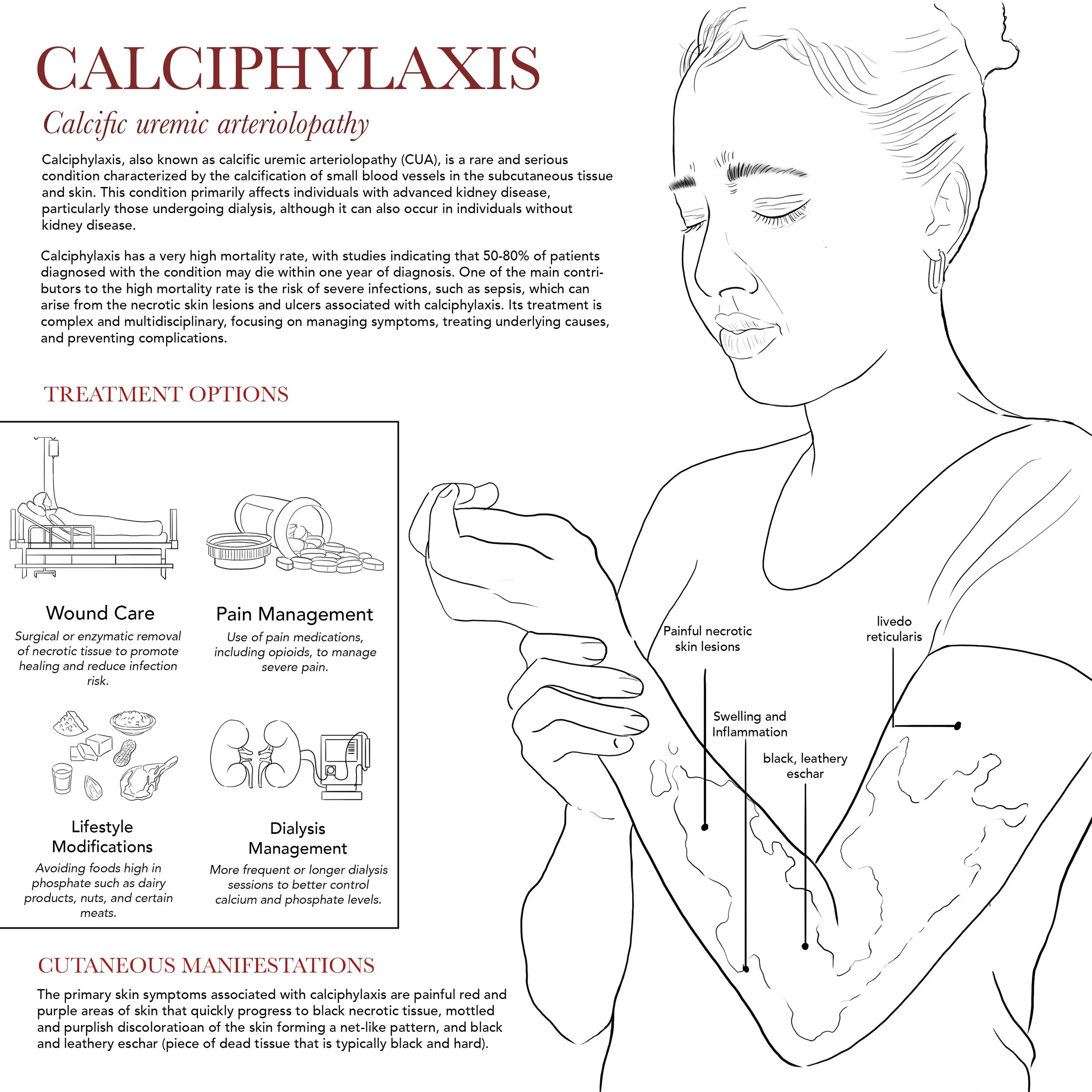
Purpose: To depict the pathology of calciphylaxis, illustrating the characteristic skin lesions with areas of necrosis and ulceration, and to show treatment options aimed at wound care, pain management, and addressing underlying metabolic imbalances.
Clinical Imagery
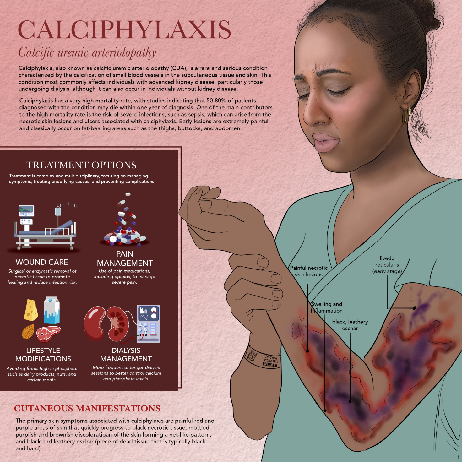
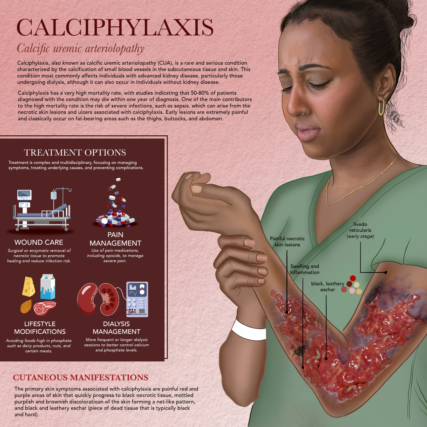
When working on this illustration, I wanted to capture the severe and debilitating nature of calciphylaxis by clearly depicting its physical appearance-painful, necrotic skin lesions with areas of ulceration. My goal was to make the visual communicate just how damaging and life-altering this condition can be. To do this accurately, I studied clinical reference images of calciphylaxis wounds to ensure I represented the skin changes realistically, including how in deeper skin tones the lesions often appear more purple along with the darker tissue necrosis. I also aimed to include key treatment approaches like wound care and pain management to give a more complete picture of how the disease is addressed.
Process Work
Ideation and Work in Progress
In developing this illustration, I focused on achieving a realistic and clinically meaningful depiction of the condition by carefully studying photographic references of skin manifestations. I wanted to emphasize the pronounced redness, swelling, and inflammation, using color and texture to convey the underlying pathology clearly. I also made sure to accurately portray livedo reticularis with its distinctive, net-like mottled pattern on the skin, highlighting how it appears in real patients.
I started with some initial sketches to play around with the composition, and as I refined the concept, I had to reconsider my subject’s appearance because calciphylaxis is such a debilitating condition and patients are often hospitalized. This led me to incorporate details like hospital clothing and a patient arm band to ground the illustration in a realistic clinical setting.
As I progressed, I slowly added in the color of the rash, carefully keeping in mind that it often appears with more purple tones in deeper skin tones to accurately reflect clinical variation. Finally, I added subtle specular highlights to indicate open wounds and areas of moist ulceration, reinforcing the severity and complexity of the condition while maintaining an illustrative quality that enhances understanding.