Conjunctivitis Infographic.
Medium: Adobe photoshop and illustrator
Client: Johnson & Johnson/ AMI
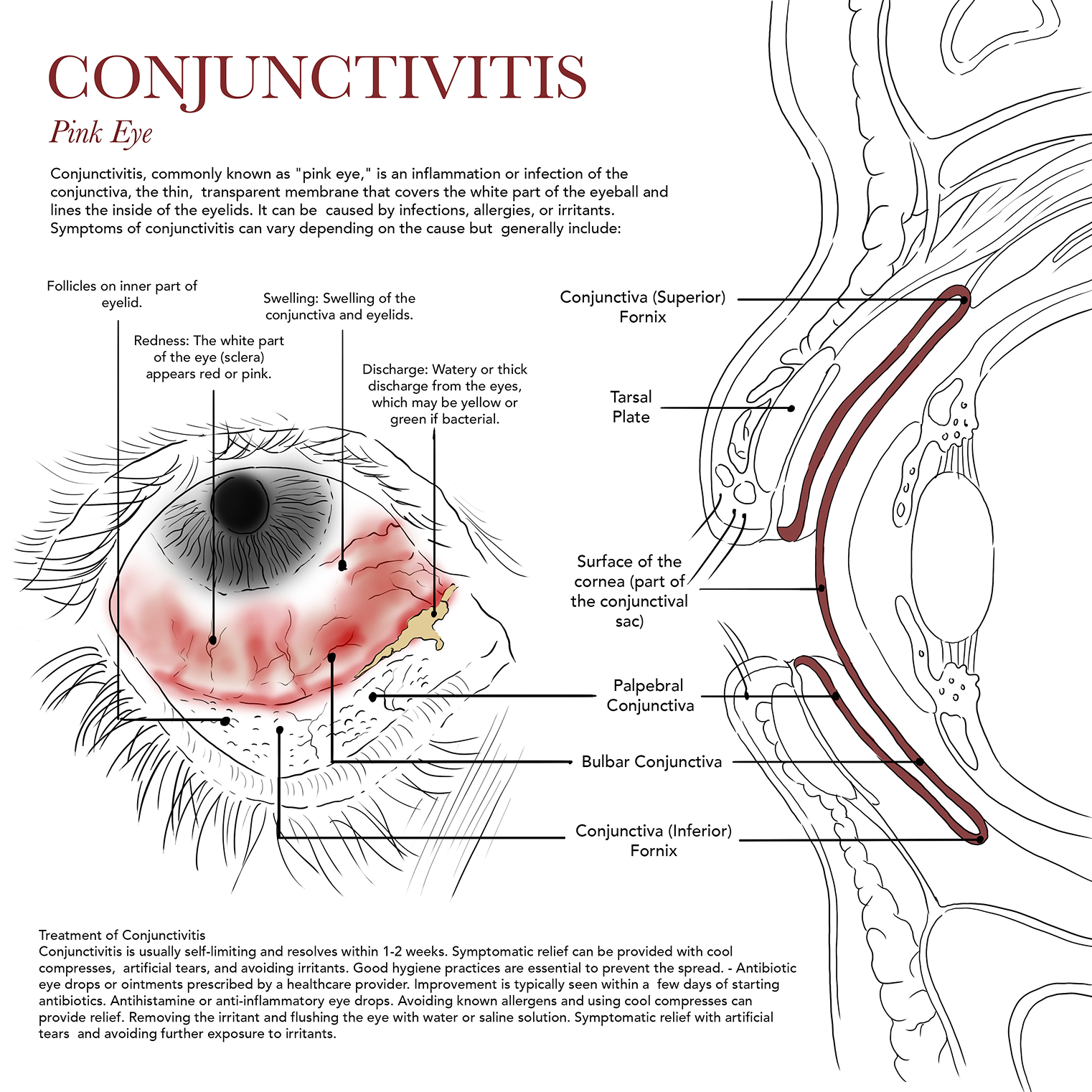
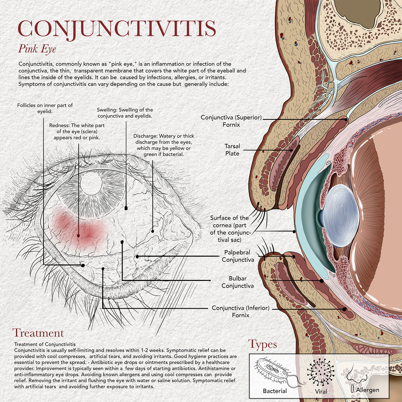
Purpose: To depict the pathology of conjunctivitis, highlighting the associated visible symptoms through both anterior and sagittal views of the eye anatomy.
Textbook and Photographic References
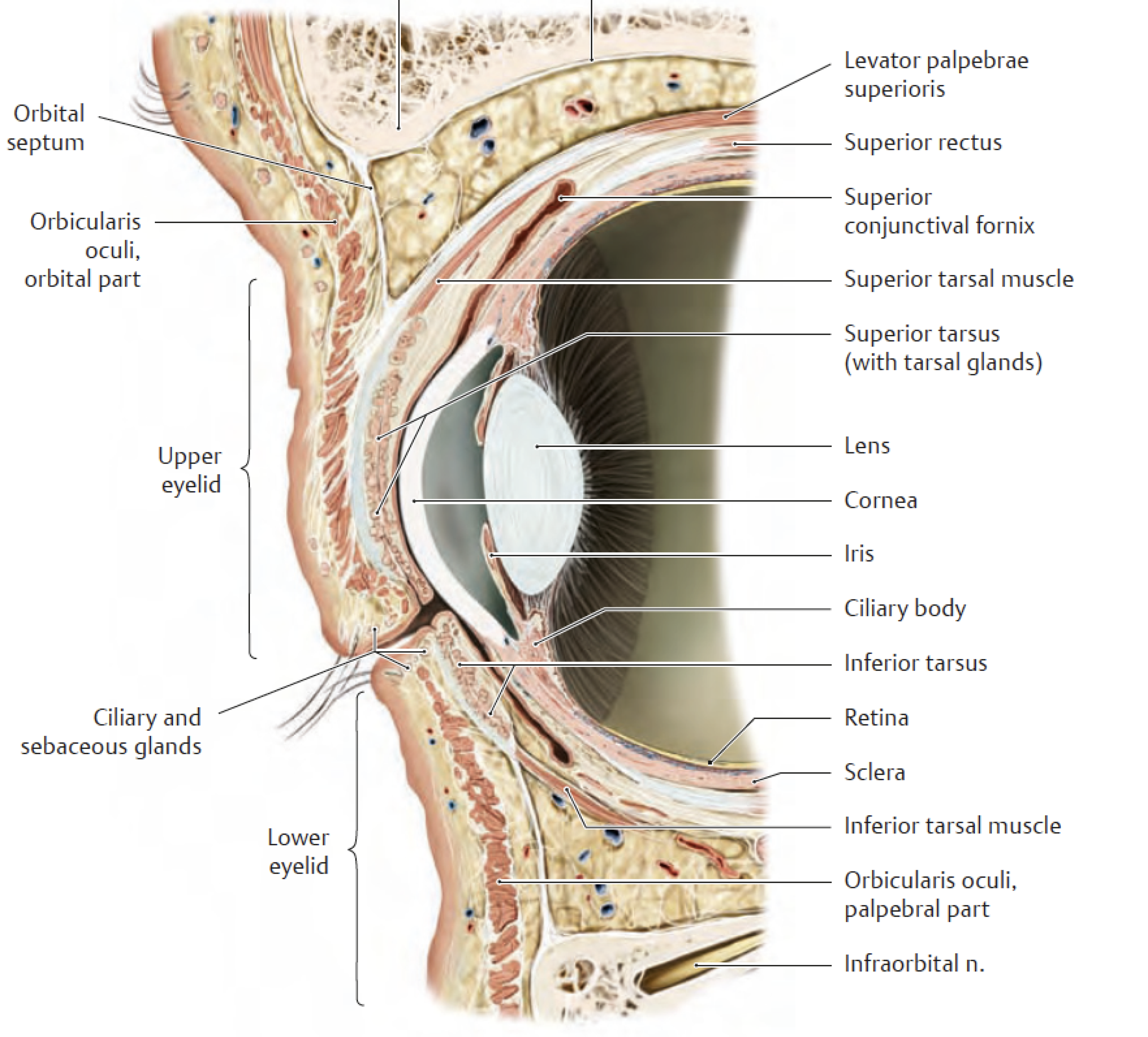
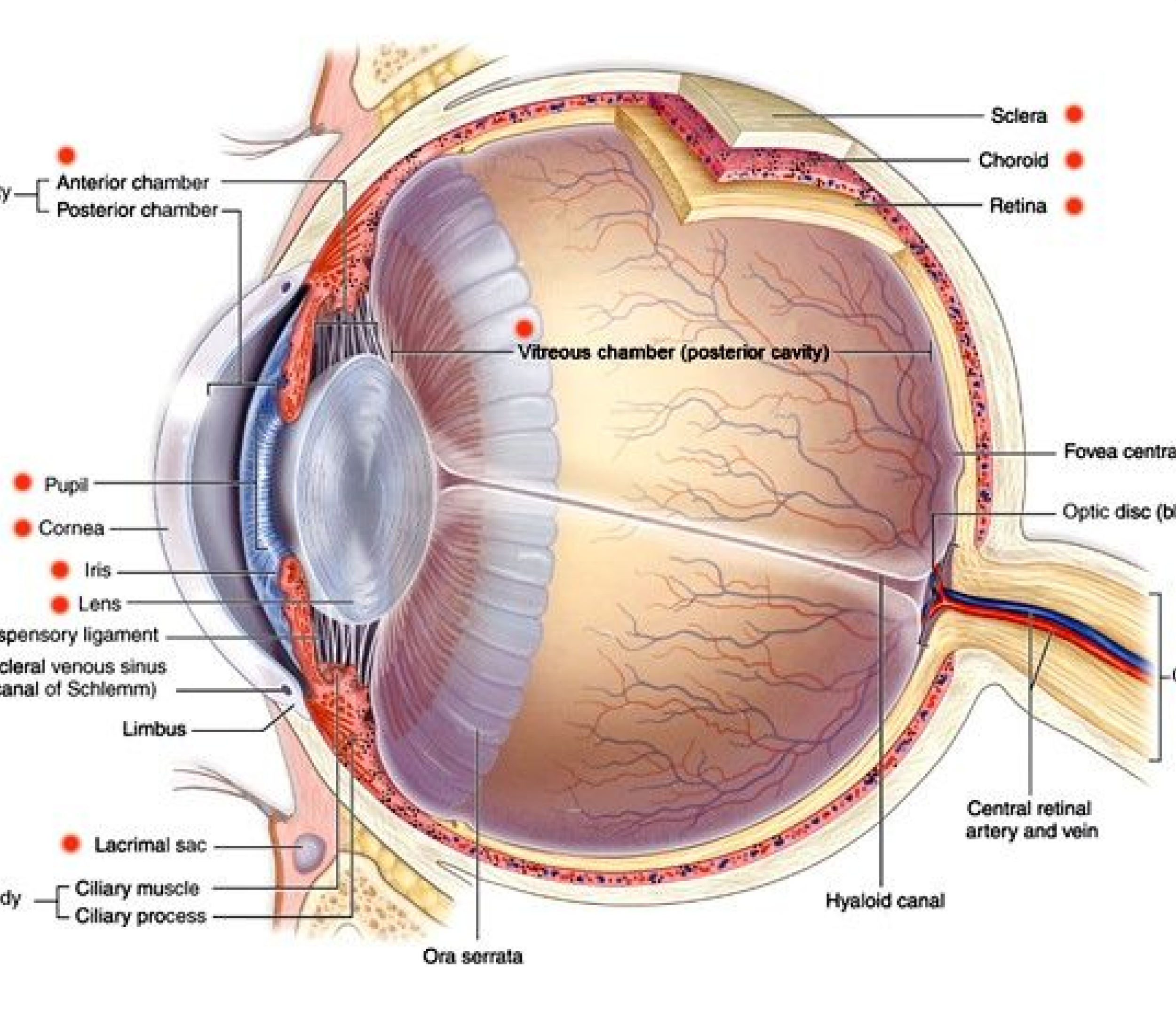
Textbook references of sagittal sections of eye anatomy were initially reviewed to ensure anatomical accuracy, supplemented by photographic references of conjunctivitis to capture its distinct features, including follicles, redness, swelling, and discharge.
Process Work

Ideation and Work in Progress
In developing this illustration, I aimed to capture a high level of anatomical detail by consulting photographic references from the NIH Image Gallery alongside ophthalmology textbook images. I also chose to retain elements of line art in the final rendering to provide a stylistic touch that enhances clarity and structure. While the sagittal cross-section of the eye was not essential for depicting conjunctivitis symptoms, I included it as an informative addition to deepen my own understanding of ocular anatomy and to illustrate how the various parts of the conjunctiva fit within this perspective.