Dermatomyositis Infographic.
Medium: Adobe photoshop and illustrator
Client: Johnson & Johnson/ AMI
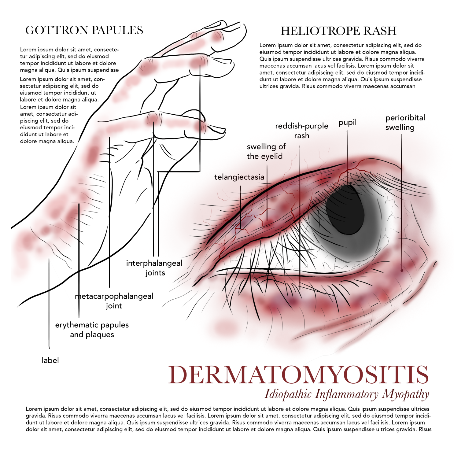
Purpose: To depict the pathology of dermatomyositis, illustrating the characteristic violaceous periorbital rash known as the heliotrope rash around the eyes, along with erythematous or violaceous, scaly papules over the knuckles (Gottron’s papules) and other photo-exposed areas.
Clinical Imagery
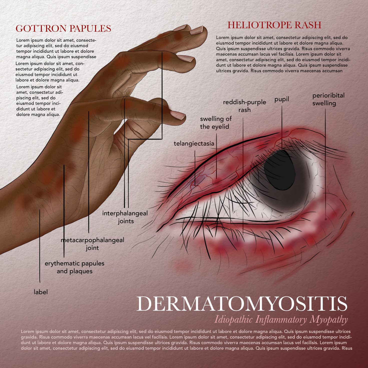
When working on this illustration, I wanted to convey the distinctive yet often subtle skin manifestations of dermatomyositis by clearly depicting its characteristic rashes-especially the heliotrope rash around the eyes and the scaly, erythematous papules over the knuckles known as Gottron’s papules. My goal was to communicate how these skin changes can be key diagnostic clues while also showing the variability in presentation. To do this accurately, I studied clinical reference images to capture how the violaceous hue of the heliotrope rash may appear less obvious in deeper skin tones, sometimes presenting with more subtle darkening or discoloration. I also aimed to give a sense of the chronic, systemic nature of the disease, suggesting the importance of early recognition and treatment with immunosuppressive therapies to manage symptoms and prevent progression.
Process Work
Ideation and Work in Progress
In developing this illustration, I wanted to create a realistic and clinically meaningful depiction of dermatomyositis by carefully studying photographic references of its characteristic skin manifestations. I chose to focus on two key areas often affected by the disease: the skin around the eyes, to highlight the heliotrope rash with its distinctive violaceous discoloration and swelling, and the hand, to show Gottron’s papules over the knuckles with their raised, scaly, inflamed appearance.
I began with initial sketches to experiment with the composition, thinking carefully about how best to capture these hallmark features in a single, cohesive visual. As I refined the concept, I considered how to clearly illustrate the underlying inflammation, using color, shading, and texture to convey redness, swelling, and subtle changes in skin surface detail.
Throughout the process, I was mindful of clinical variation across skin tones, recognizing that the heliotrope rash can appear less vivid in deeper skin tones-sometimes presenting as darker or more muted discolouration rather than bright purple. I worked to represent this variation accurately to make the illustration inclusive and useful for recognizing dermatomyositis in diverse patient populations. As I layered in color and detail, I focused on maintaining a balance between anatomical accuracy and an illustrative quality that would help viewers clearly understand the nature of the inflammation and its characteristic distribution.