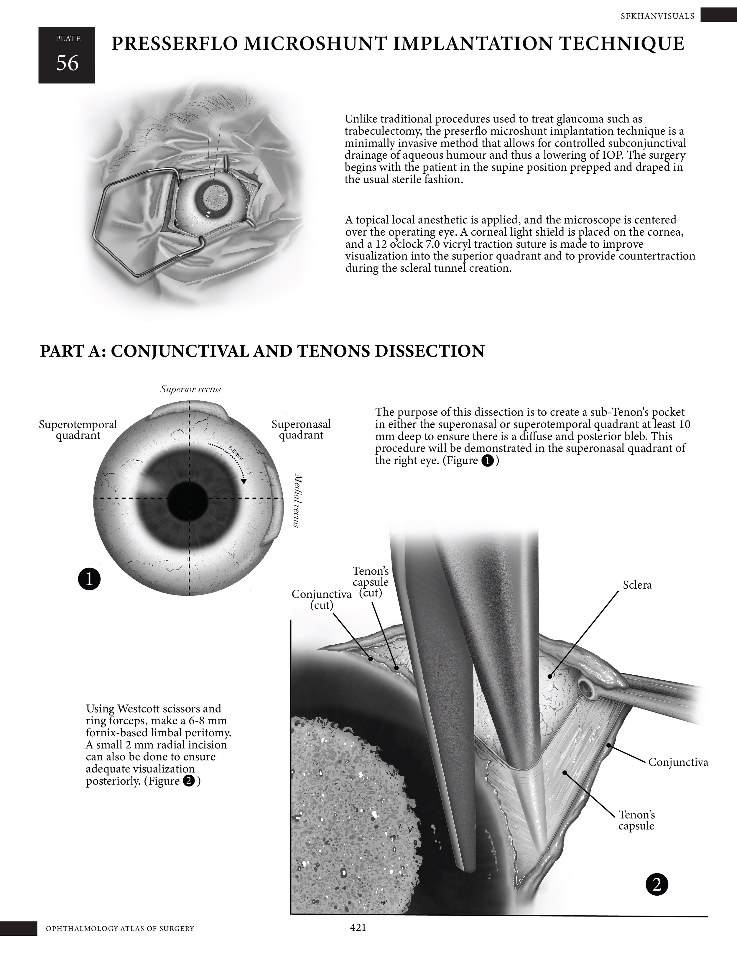
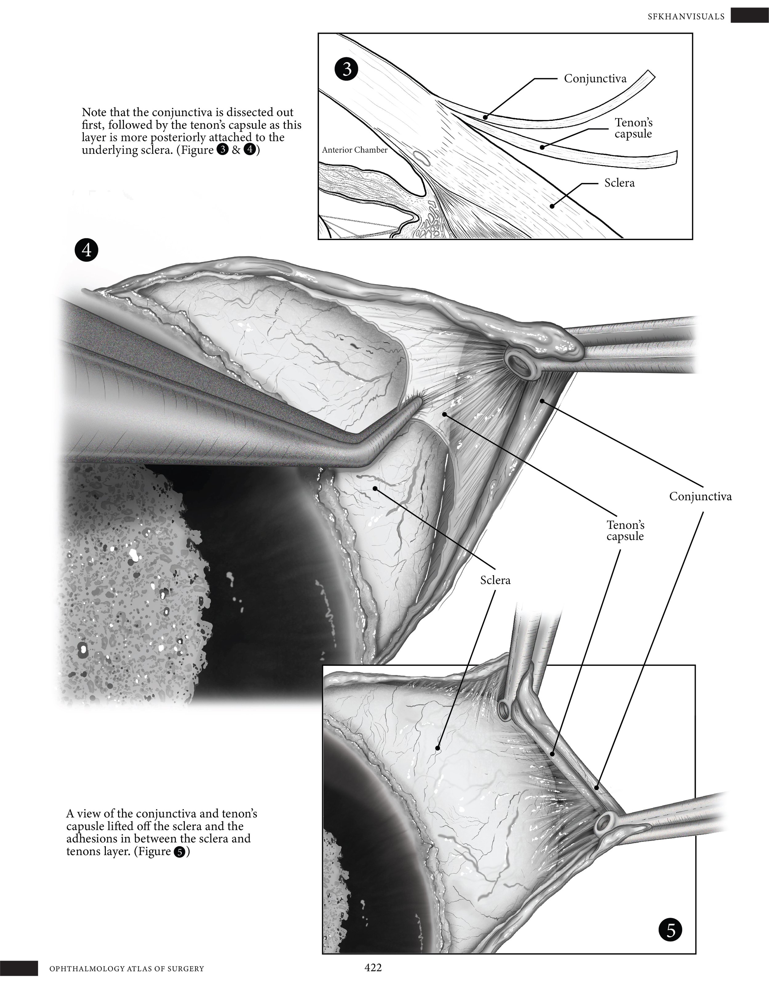
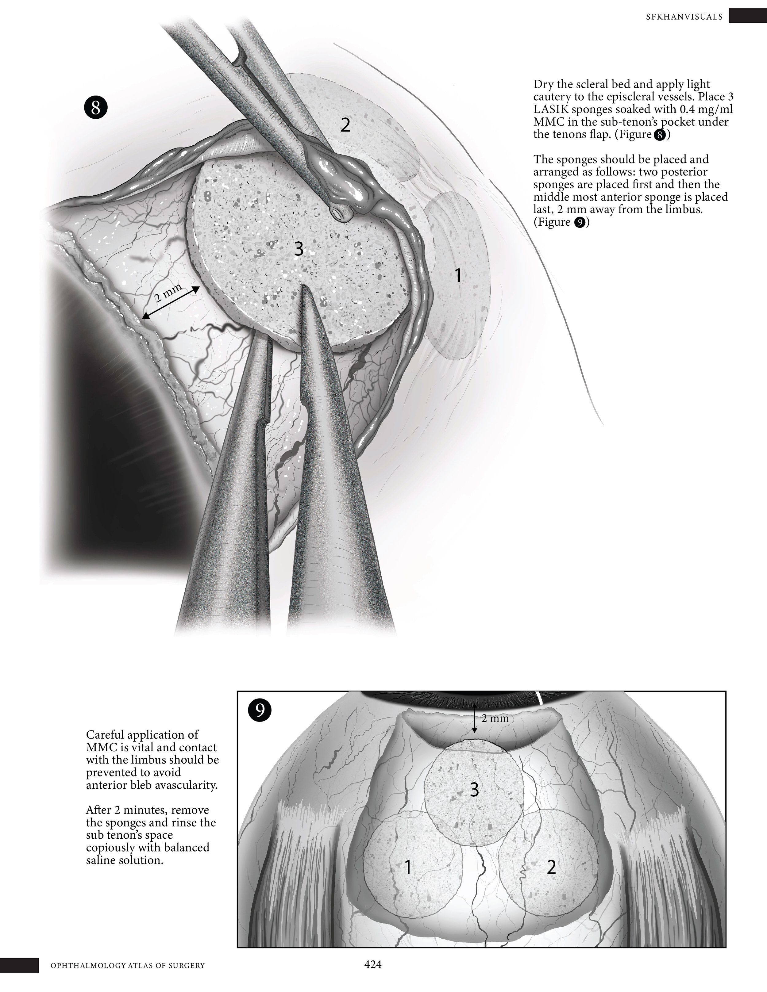
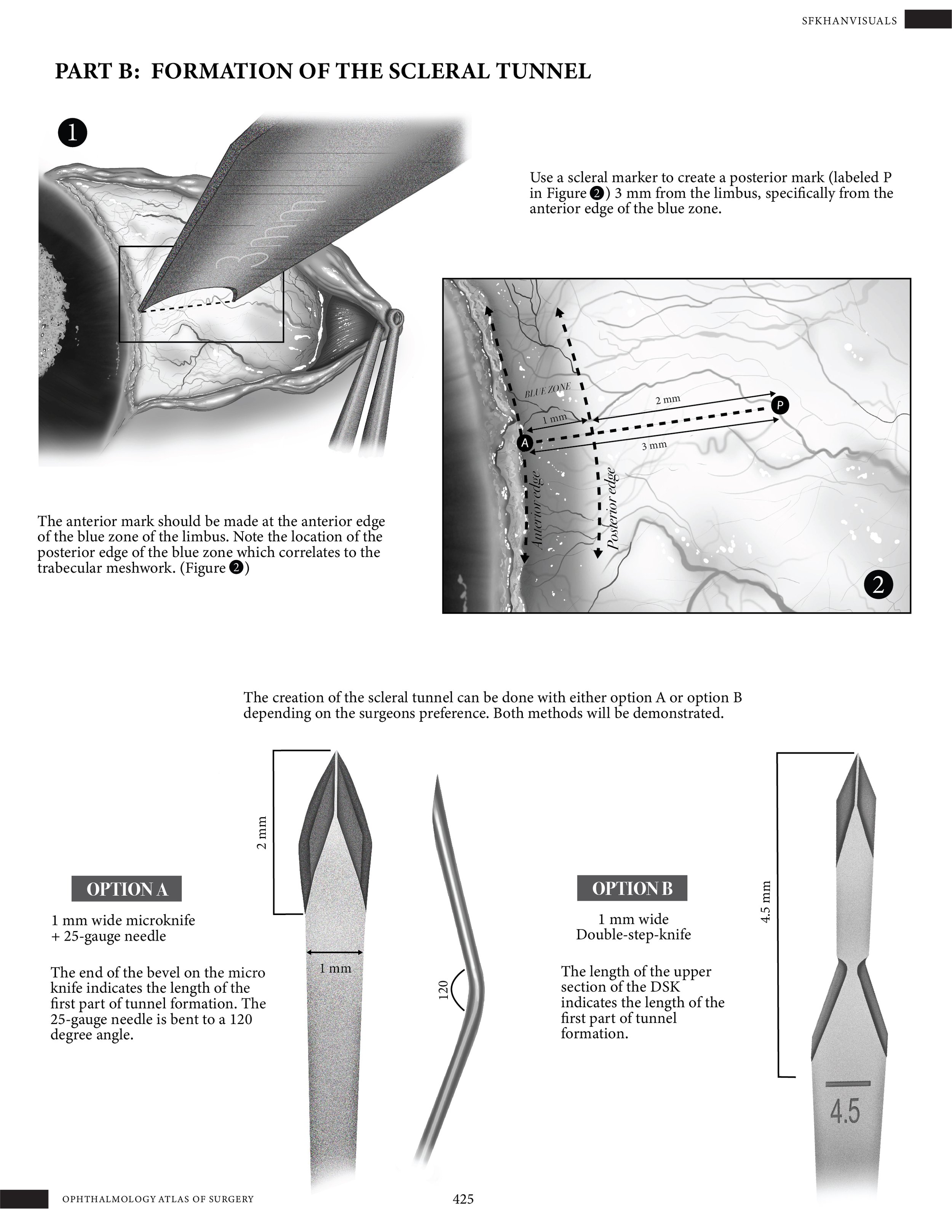
Preserflo Microshunt Implantation Surgical Guide
Medium: Adobe photoshop and illustrator
Client: Dr. Ike Ahmed, Dr. Ema Avdagic & Dr. Ticiana De Francesco (Credit Valley Hospital)
Purpose: To illustrate the surgical sequence involved in the implantation of a Preserflo microshunt intended to reduce intraocular pressure within the eye.
Operating Room Surgical Sketches
Preliminary surgical sketches were created on-site in the operating room while observing the procedure being performed on multiple patients. Working alongside the surgical team, I captured key stages of the operation in real time, focusing on anatomical accuracy, surgical technique, and the positioning of instruments. These sketches served as valuable visual references for understanding procedural flow and informed the development of more refined illustrations post-operatively. The dynamic environment required quick observation, adaptability, and a strong foundation in anatomical knowledge to effectively document each step while maintaining respect for the sterile field and patient privacy.
Process Work
Development of Reference Boards
To support the development of the final illustration, I created multiple reference boards tailored to different aspects of the visualization process. The first board focused on rendering inspiration, compiling examples of stylistic approaches, lighting, and texture treatments to guide the overall aesthetic direction. The second board featured anatomical references, including textbook illustrations and cadaveric imagery, to ensure structural accuracy and proper spatial relationships between key anatomical landmarks. The third board was dedicated to surgical references, showcasing intraoperative photos and procedural diagrams to capture specific details of the technique and instrumentation.
In addition to static references, I was given access to patient surgical videos provided by the content advisors. These videos offered dynamic, real-time insights into the procedure, allowing me to observe subtle tissue interactions, surgeon hand positioning, and the step-by-step progression of the surgery.
Development of the First Draft
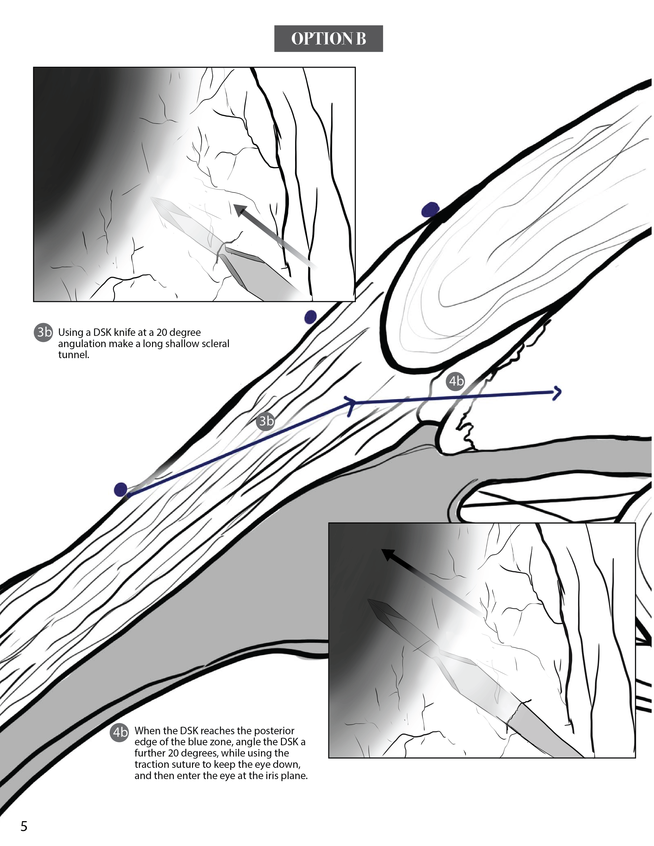
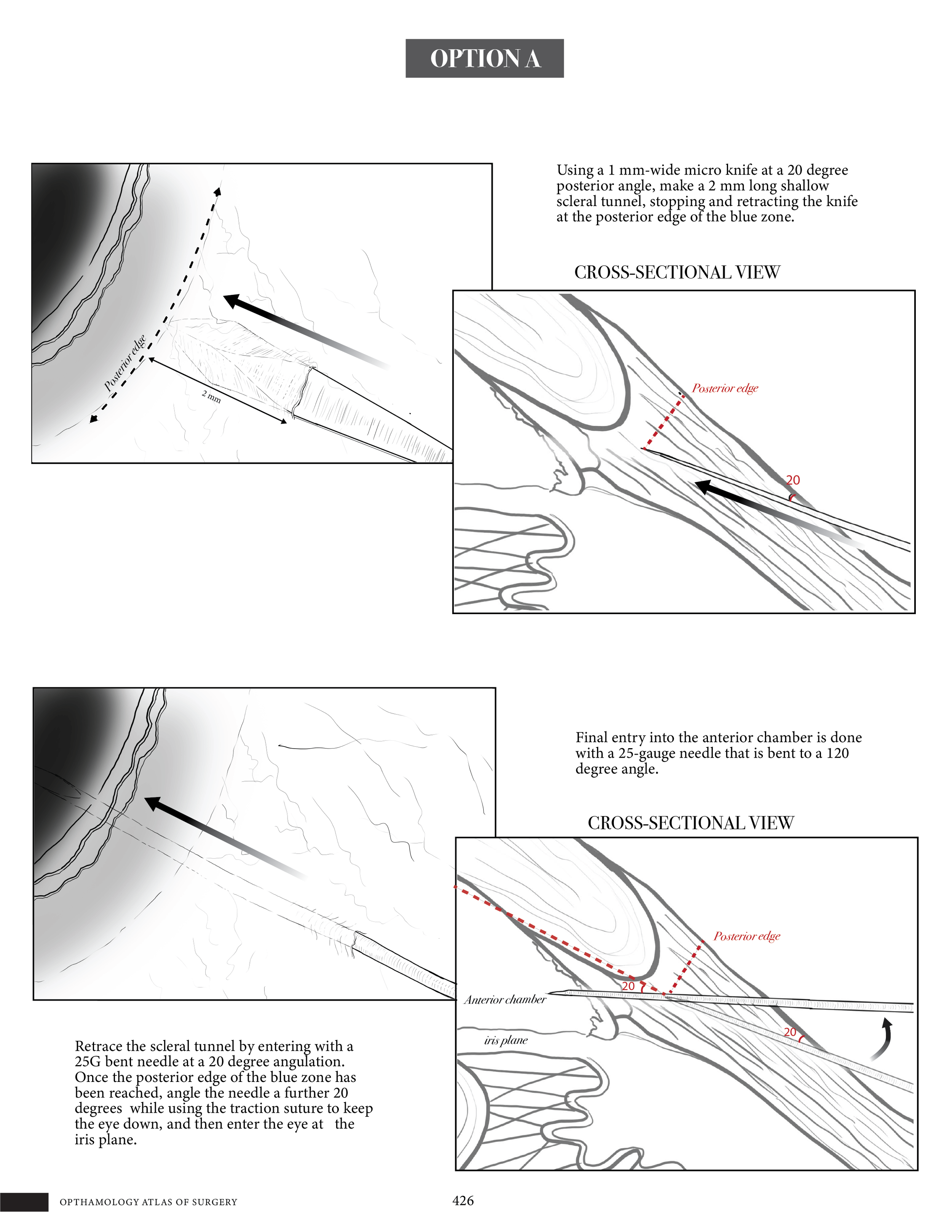
Given the complexity and length of the procedure, I organized the surgical process into clearly defined sections labeled A through B, with each section further broken down into multiple detailed steps. This structured approach helped to streamline the visualization of the procedure and made it easier to follow the surgical narrative in a logical and sequential manner.
Because the final illustration was intended as instructional material for surgical residents, clarity and pedagogical value were key priorities. To support this, I maintained a consistent viewpoint that closely mirrored the surgeon’s perspective during the operation. This decision allowed for a more intuitive understanding of hand positioning, instrument use, and anatomical orientation-ultimately enhancing the educational value of the illustration by aligning it with what residents would observe in the operating room.


Development of the Second Draft
Following a detailed review of the initial draft by the content advisors, a refined version of the illustration was developed with a stronger emphasis on anatomical accuracy and surgical precision. Feedback from subject matter experts guided several key adjustments, including the correction of spatial relationships between anatomical structures and improved depiction of tissue planes and surgical landmarks.
To enhance clarity and instructional value, graphical elements were thoughtfully integrated into the refined version. Leader lines were used to connect labels directly to relevant anatomical features without crowding the image, while callout boxes provided additional context for critical steps or complex structures. Directional arrows were incorporated to indicate the motion of instruments, incision paths, or procedural flow, helping to guide the viewer through the sequence more intuitively.
The overall layout was carefully finalized to ensure a logical visual progression through the surgical steps, maintaining a balance between detail and readability. This final composition served as a clear, accurate, and accessible teaching tool for surgical trainees.


Development of Finished Rendered Draft
The final draft was rendered in grayscale using Adobe Photoshop to achieve both anatomical accuracy and a high degree of realism, specifically tailored to the complex structures of the eye. The grayscale approach allowed for precise control over values, enabling the careful depiction of delicate ocular tissues such as the sclera, cornea, iris, and surrounding musculature without the visual distraction of color.